Shockwave Therapy for Plantar Fasciitis in Murray, UT: The Anatomy, the Causes, and How to Get Back on Your Feet

If your first steps in the morning feel like stepping on a nail, you are not alone. Plantar fasciitis is one of the most common causes of heel pain, affecting roughly one in ten people at some point in their lives. Most patients who walk or run into our Murray, Utah, sports chiropractic office have already tried rest, new shoes, and stretching, only to find the pain keeps coming back or really never left. The good news is that heel pain is highly treatable once you understand what is actually happening in the tissue, what actions can help you improve and get back to your favorite activities, and where extracorporeal shockwave therapy (ESWT) fits in the care plan.

The Anatomy: What Is the Plantar Fascia?

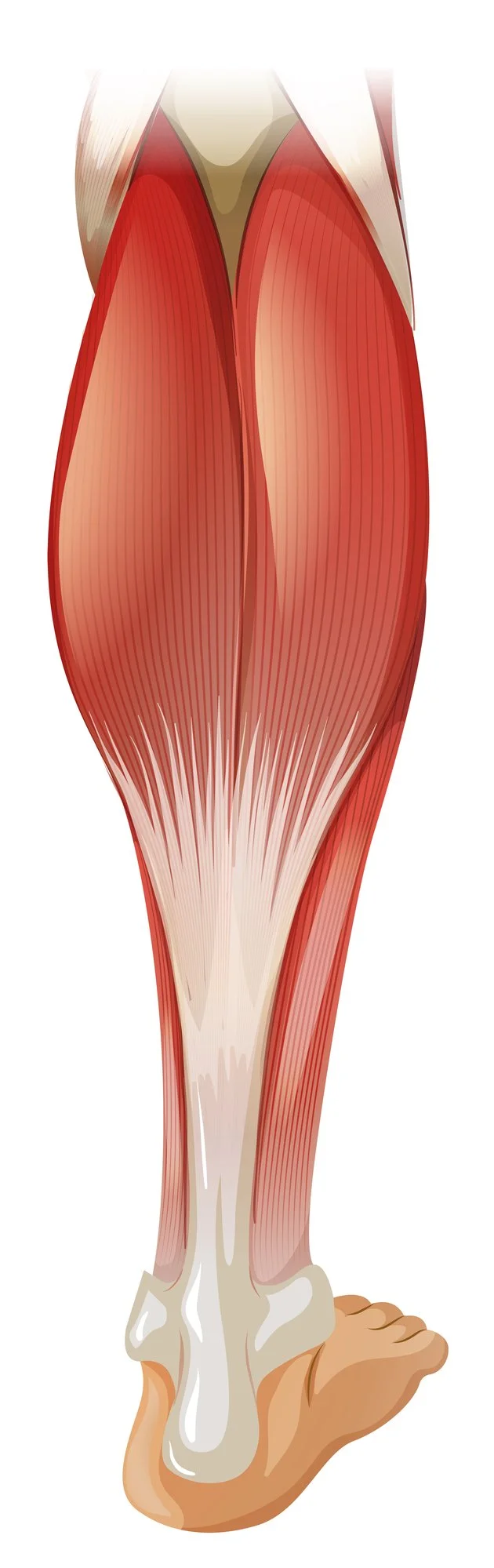
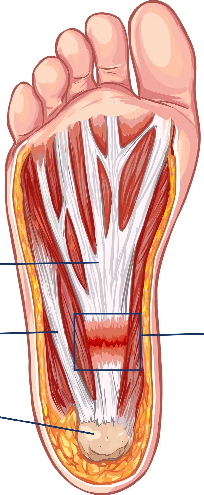
Plantar fascia anatomy and how shockwave therapy can help decrease pain and heal damage.

Before we start to attack this common issue, knowing the anatomy involved is important. The plantar fascia is a thick, fibrous band of connective tissue that runs along the sole of your foot, connecting your heel bone (the calcaneus) to the base of your toes. Think of it as a strong bowstring that supports the arch of your foot and absorbs shock every time your foot strikes the ground. When you walk, run, or jump, this tissue stretches and recoils thousands of times a day, storing and releasing energy to make each step efficient.


Despite the "-itis" in its name, plantar fasciitis is now understood to be less about acute inflammation and more about degeneration and disorganized tissue repair. Under repeated overload, tiny tears form where the fascia attaches to the heel, and the body's healing response falls behind the damage. Because this is a degenerative process rather than a simple inflammatory one, researchers increasingly use the term "plantar fasciopathy." [2] That distinction matters because it explains why anti-inflammatory measures alone often fail and why treatments that stimulate healing and increase tissue capacity tend to work better.

test


Common Causes of Plantar Fasciitis

Plantar fasciitis develops when the repetitive load on the fascia outpaces its ability to recover. Common contributors include a sudden increase in running or walking mileage, spending long hours on your feet on hard surfaces, tight calf muscles and Achilles tendons that limit ankle mobility, flat feet or high arches that change how load is distributed across the foot, unsupportive footwear, and carrying extra body weight. Age plays a role too, with cases peaking between 40 and 60 years old. Often it is not one factor but several stacking together over weeks or months.

How Plantar Fasciitis Limits Your Favorite Activities

The hallmark of plantar fasciitis is sharp, stabbing heel pain with your first steps in the morning or after sitting for a while, which may ease as you warm up and then return later in the day. For active people, the impact goes far beyond discomfort. Runners find their mileage cut short or their training halted entirely. Hikers hesitate before tackling the trails in the Wasatch. Parents, nurses, teachers, and tradespeople who are on their feet all day dread the end-of-shift ache. Many people unconsciously alter their gait to avoid the pain, which can trigger new problems in the knees, hips, and lower back. Left unaddressed, plantar fasciitis can linger for a year or more, quietly eroding the activities that keep you healthy and happy.

Why Shockwave Therapy Is a Leading Option for Plantar Fasciitis Relief

When rest, stretching, and orthotics have not solved the problem, extracorporeal shockwave therapy has emerged as one of the best-supported non-surgical treatments for chronic plantar fasciitis. ESWT delivers focused acoustic pressure waves into the affected tissue. These mechanical pulses are thought to work by stimulating new blood vessel formation (neovascularization), triggering collagen production and tissue remodeling, and disrupting the pain signals that keep the cycle going, essentially prompting a stalled healing process to restart.

Shockwave Therapy for Plantar Fasciitis Relief

The evidence is strong. In a landmark randomized, placebo-controlled, multicenter trial, Gerdesmeyer and colleagues found that shockwave therapy produced significantly greater improvements in heel pain and function than placebo in patients with chronic recalcitrant plantar fasciitis. [1] Multiple systematic reviews and meta-analyses of randomized controlled trials have since confirmed that ESWT is both effective and safe for chronic plantar fasciitis. [3] [4] Compared with corticosteroid injections, shockwave delivers similar or better relief without the risk of fascia thinning or rupture that comes with repeated steroids. [5]

Importantly, shockwave works best as part of a comprehensive plan. At Revive Sport & Spine, ESWT is combined with progressive loading exercises, targeted calf and plantar fascia stretching, and gait retraining. The research is consistent on this point: addressing the underlying tissue capacity through loading, alongside shockwave to jump-start healing, gives the most durable results, far greater than rest alone. [4]

What to Expect with Shockwave Therapy for Plantar Fasciitis

A typical shockwave session lasts about 5 to 15 minutes, requires no anesthesia, and lets you walk out the door immediately. Most patients complete a series of 4 to 8 sessions spaced 7 to 10 days apart, with continued improvement often seen up to 12 weeks after treatment as the tissue remodels. You may feel a deep tapping or mild discomfort during the session, and the intensity is adjustable to your comfort. To let your body's natural healing response do its work, avoid anti-inflammatory medication (as directed by your PCP) and icing for 48 hours after each session.

Take the First Step Toward Relief from Plantar Fasciitis

You do not have to let heel pain sideline your runs, hikes, and daily life. Call Revive Sport & Spine at 801-944-1855 or schedule online to find out whether shockwave therapy is right for you. We are located at 1996 E 6400 S, Suite 260, in Murray, Utah, proudly serving Salt Lake City, Holladay, Midvale, Sandy, Cottonwood Heights, Millcreek, and the greater Salt Lake Valley.


Frequently Asked Questions

Does shockwave therapy for plantar fasciitis hurt? Most patients describe it as a tolerable deep tapping sensation. Intensity is adjusted to your comfort, and there is no downtime afterward.

How many sessions will I need? Most people benefit from 4 to 8 sessions, though your exact plan depends on how long you have had symptoms and how you respond.

Is it better than a cortisone shot? Research shows shockwave delivers comparable or better long-term relief without the risk of fascia thinning or rupture associated with repeated steroid injections. [5]


References

[1] Gerdesmeyer L, Frey C, Vester J, et al. Radial extracorporeal shock wave therapy is safe and effective in the treatment of chronic recalcitrant plantar fasciitis: results of a confirmatory randomized placebo-controlled multicenter study. The American Journal of Sports Medicine. 2008;36(11):2100-2109. https://pubmed.ncbi.nlm.nih.gov/18832341/

[2] Rompe JD, Furia J, Weil L, Maffulli N. Shock wave therapy for chronic plantar fasciopathy. British Medical Bulletin. 2007;81-82:183-208. https://pubmed.ncbi.nlm.nih.gov/17456546/

[3] Sun J, Gao F, Wang Y, Sun W, Jiang B, Li Z. Extracorporeal shock wave therapy is effective in treating chronic plantar fasciitis: a meta-analysis of RCTs. Medicine (Baltimore). 2017;96(15):e6621. https://pubmed.ncbi.nlm.nih.gov/28403111/

[4] Lou J, Wang S, Liu S, Xing G. Effectiveness of extracorporeal shock wave therapy without local anesthesia in patients with recalcitrant plantar fasciitis: a meta-analysis of randomized controlled trials. American Journal of Physical Medicine & Rehabilitation. 2017;96(8):529-534. https://pubmed.ncbi.nlm.nih.gov/27977431/

[5] Xiong Y, Wu Q, Mi B, et al. Comparison of efficacy of shock-wave therapy versus corticosteroids in plantar fasciitis: a meta-analysis of randomized controlled trials. Archives of Orthopaedic and Trauma Surgery. 2019;139(4):529-536. https://pubmed.ncbi.nlm.nih.gov/30426211/

Knee Osteoarthritis: Risk Factors, Prevention, Nutrition, and When to Seek Care

Knee osteoarthritis is one of the most common reasons people walk into our sports chiropractic office in Murray, and it is also one of the most misunderstood. In Part 1, we described knee osteoarthritis (OA) as a whole-joint condition driven by a mismatch between joint load and its capacity to handle it. That framing is useful because it points directly to what you can influence. This article covers the factors that raise your risk, the strategies that genuinely help with prevention, what the nutrition research does and doesn’t support, and the signs that mean it’s time to have your knee looked at.

What Raises Your Risk of Knee Osteoarthritis?

Some risk factors are outside your control. Age increases risk; women are affected more often than men; genetics play a role; and a previous significant knee injury, such as an ACL tear, meniscus damage, or fracture, raises the odds of developing OA in that joint years later. Joint alignment and anatomy matter too.

The good news is that several of the most powerful risk factors are modifiable. Body weight is at the top of that list. Excess weight increases the mechanical load through the knee with every step, but it also contributes through metabolic and inflammatory pathways, which is why obesity is linked to OA even in non-weight-bearing joints. Research consistently shows that higher body weight is associated with both a greater risk of developing knee OA and more severe pain and functional limitation once it is present. 1 6 Other modifiable contributors include weak or deconditioned muscles around the knee and hip, occupations or activities involving heavy repetitive loading, and poorly managed prior injuries.

A Quick Anatomy Review

Between the femur and tibia sit two C-shaped shock absorbers called the menisci. They spread the load across the joint, add stability, and protect the cartilage underneath. Surrounding the whole joint is a capsule lined with synovium, a thin membrane that produces synovial fluid to lubricate and nourish the cartilage. Ligaments such as the ACL, PCL, LCL, and MCL hold the bones in alignment, while the muscles that cross the knee, especially the quadriceps and hamstrings, control movement and absorb force. Just beneath the cartilage lies the subchondral bone, which plays a bigger role in osteoarthritis than most people realize.

What Actually Helps to Prevent Osteoarthritis (or slows it down)

Because weight is such a strong and modifiable driver, weight management is one of the highest-yield prevention strategies available. A meta-analysis of observational studies estimated that preventing obesity could meaningfully reduce the incidence of knee osteoarthritis at the population level by almost 30. 2 For someone already carrying extra weight, even modest weight loss reduces knee load and symptoms.

Staying active is the second pillar. Contrary to the old fear that exercise “wears out” the knees, regular moderate activity and strong supporting muscles protect the joint by improving its capacity to absorb load. Keeping the quadriceps, hamstrings, and hip muscles strong, maintaining good range of motion, and addressing prior injuries properly all help keep the load-to-capacity balance in your favor. We will cover the specifics of exercise in Part 4 of this series.

How Can Your Nutrition Affect Your Risk of Osteoarthritis?

Patients often ask which foods or supplements will fix their knees. It’s worth being honest here: no diet or supplement reverses established osteoarthritis, and the marketing around joint supplements far outpaces the evidence. That said, nutrition is not irrelevant. 3

The strongest dietary evidence supports an overall healthy, anti-inflammatory eating pattern. Something similar to a Mediterranean-style diet rich in vegetables, fruit, whole grains, fish, and healthy fats, and lower in ultra-processed foods and added sugar. Long-chain omega-3 fatty acids (from oily fish) and adequate vitamin D are frequently discussed, and correcting a genuine deficiency is reasonable, but evidence that isolated supplementation alters the disease course is limited. 3 4 Popular products like glucosamine and chondroitin have mixed reviews, with some patients having good relief with consistent use, but major studies and guidelines do not strongly recommend them. 5

The practical takeaway: use nutrition to manage weight and support overall health rather than chasing a single “joint miracle” supplement. If you’re considering supplements, it’s worth discussing them with your healthcare team to find the best options for your particular concerns.

When Should You Seek Care for Your Knee Pain?

Knee pain is easy to dismiss, but certain signs mean you shouldn’t wait. Consider getting your knee evaluated if you have pain that persists for more than a few weeks, pain that limits walking, climbing stairs, work, or sleep, ongoing swelling, a knee that locks, catches, or gives way, or stiffness that consistently lingers after rest.

NOTE: Rapidly worsening pain, a hot and visibly swollen joint, fever, or an inability to bear weight warrants prompt medical attention, as those can signal something other than routine osteoarthritis.

Seeking care early has a real advantage: the sooner you address strength, movement, and load, the more you can do to keep the joint functioning well and avoid the deconditioning spiral described in Part 1. Guidelines recommend starting with conservative, active management rather than jumping to imaging or surgery for most people. 5

In Part 3, we’ll walk through exactly how we care for knee osteoarthritis at Revive Sport & Spine, including soft-tissue therapyjoint manipulation, and shockwave therapy, and how these approaches set the stage for the rehab exercise plan in Part 4.

Key Takeaways Before We Progress to Part 3

✓  Excess body weight through diet and exercise is one of the strongest and most modifiable risk factors for knee osteoarthritis. 1 2

✓  No supplement “cures” OA, but weight management and an overall healthy, anti-inflammatory dietary pattern have the best evidence. 3 4

✓  Persistent swelling, locking, giving way, or pain that limits daily life are signals to get your knee evaluated. 5

If your knee has been nagging or you have risk factors and want to stay ahead of osteoarthritic changes and pain, an assessment is a smart first step. We’ll assess your movement, strength, and daily load and help you develop a realistic prevention or management plan.


Frequently Asked Questions

What is the biggest risk factor for knee osteoarthritis?

Excess body weight is among the strongest and most modifiable risk factors. It increases mechanical load on the knee, drives inflammation, and is linked to both developing OA and worse symptoms. 1 2 6

Can knee osteoarthritis be prevented?

You can’t change age or genetics, but you can lower your risk by maintaining a healthy weight, staying active, keeping the muscles around the knee strong, and properly rehabilitating any knee injuries. Preventing obesity alone could reduce up to 30% of knee OA cases. 2

What is the best diet for knee osteoarthritis?

There is no magic food, but the best-supported approach is using nutrition to reach and maintain a healthy weight alongside an overall anti-inflammatory, Mediterranean-style pattern rich in vegetables, fish, and healthy fats. 3 4

Do glucosamine and chondroitin work for knee arthritis?

While some patients swear by it, high-quality trials show inconsistent results, and major clinical guidelines do not strongly recommend them. They are generally safe, but they should not replace weight management, exercise, a reasonable diet, and appropriate care. 5

When should I see someone about knee pain?

Get evaluated if pain lasts more than a few weeks, limits daily activities, or is accompanied by swelling, locking, or giving way. A hot, swollen joint with fever or an inability to bear weight needs prompt emergency medical attention. 5


REFRENCES

1. Batushansky A, et al. Obesity-Related Knee Osteoarthritis — Current Concepts. (Review). 2023. https://pmc.ncbi.nlm.nih.gov/articles/PMC10456094/

2. Zheng H, Chen C. What if we prevent obesity? Risk reduction in knee osteoarthritis estimated through a meta-analysis of observational studies. Arthritis Care & Research. 2011. https://pubmed.ncbi.nlm.nih.gov/21425246/

3. Diet in Knee Osteoarthritis — Myths and Facts. Nutrients. 2025;17(11):1872. https://www.mdpi.com/2072-6643/17/11/1872

4. Thomas S, et al. What is the evidence for a role for diet and nutrition in osteoarthritis? Rheumatology. 2018;57(suppl_4):iv61-iv74. https://academic.oup.com/rheumatology/article/57/suppl_4/iv61/4975692

5. Bannuru RR, et al. OARSI guidelines for the non-surgical management of knee, hip, and polyarticular osteoarthritis. Osteoarthritis and Cartilage. 2019;27(11):1578-1589. https://pubmed.ncbi.nlm.nih.gov/31278997/

6. Onishi K, et al. Level of obesity is directly associated with the clinical and functional consequences of knee osteoarthritis. Scientific Reports. 2020;10:3601. https://www.nature.com/articles/s41598-020-60587-1

Understanding Knee Osteoarthritis: The Anatomy and How It Actually Develops

Knee osteoarthritis is one of the most common reasons people walk into our sports chiropractic office in Murray, and it is also one of the most misunderstood. Most patients arrive believing their knee is simply “worn out” and that nothing can be done but wait for a replacement. The reality is more hopeful and more interesting. To make good decisions about your knee, it helps to understand what the joint is made of and what is actually happening inside it when osteoarthritis develops. That is the goal of this first article in our four-part series.

A Quick Tour of the Knee Joint - Involved Anatomy

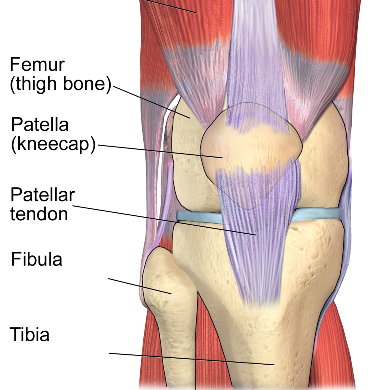
The knee is where three bones meet: the femur (thigh bone), the tibia (shin bone), and the patella (kneecap). Where these bones meet, their ends are capped with articular cartilage, a smooth, slippery, rubbery tissue that is only a few millimeters thick. Healthy cartilage is remarkably low-friction, allowing the bones to glide across one another thousands of times a day without any pain or grinding.

Between the femur and tibia sit two C-shaped shock absorbers called the menisci. They spread load across the joint, add stability, and protect the cartilage underneath. Surrounding the whole joint is a capsule lined with synovium, a thin membrane that produces synovial fluid to lubricate and nourish the cartilage. Ligaments such as the ACL, PCL, LCL, and MCL hold the bones in alignment, while the muscles that cross the knee, especially the quadriceps and hamstrings, control movement and absorb force. Just beneath the cartilage lies the subchondral bone, which plays a bigger role in osteoarthritis than most people realize.

So, What is Osteoarthritis, Really?

For decades, osteoarthritis was described as “wear and tear” of the knee joint, as if the joint were a tire slowly balding. Current research paints a very different picture. OA is now understood as an active disease of the entire joint, not just passive erosion of cartilage. Cartilage, subchondral bone, the synovium, ligaments, and even the surrounding muscles are all involved in the process. 1

In a healthy joint, cartilage is constantly maintained by cells called chondrocytes, which balance the breakdown and rebuilding of the cartilage matrix. In osteoarthritis, that balance tips toward breakdown. Chondrocytes shift into a more inflammatory, catabolic state, releasing enzymes that degrade the cartilage faster than it can be repaired. Low-grade inflammation in the joint lining adds to the problem, and the subchondral bone begins to remodel and stiffen, sometimes forming the bony spurs (osteophytes) seen on X-rays. 1 2


Why the Joint can Hurt, and Why it Sometimes Doesn’t.

Here is a detail that surprises many patients: cartilage itself has no nerve endings and no blood supply. That means early cartilage changes can be completely painless. Pain in osteoarthritis usually arises from other structures, such as the richly innervated subchondral bone, the inflamed synovium, the stretched joint capsule, and the irritated surrounding tissues. 23 This is one reason the amount of change on an X-ray often does not match the amount of pain a person feels. Some people with dramatic imaging have little pain, while others with mild imaging changes experience considerable pain.

What Drives the Knee Osteoarthritic Process Forward?

Osteoarthritis is best understood as an imbalance between the load placed on a joint and that joint’s ability to handle it. Mechanical stress is a key driver: repetitive overload, previous injury, joint malalignment, or weak supporting muscles can all push the joint past what it can comfortably tolerate, triggering the cellular and inflammatory changes described above. 1 Once the cycle starts, pain leads to less movement, less movement leads to weaker muscles and stiffer tissue, and a more poorly supported joint is loaded even less evenly, which can accelerate the degenerative process.

That cycle is exactly why the modern, evidence-based approach to knee OA emphasizes restoring the joint’s capacity through movement, strengthening, and load management rather than simply resting and waiting. International treatment guidelines now list exercise and appropriate physical care as core, first-line strategies for knee osteoarthritis. 4

Key Takeaways Before we Progress to Part 2

Knee osteoarthritis is a whole-joint condition driven by a mismatch between load and capacity, not an inevitable, untreatable breakdown. Because the joint is a living, adaptable system, the way you load it, strengthen it, and care for it genuinely matters. In Part 2, we’ll look at the specific factors that raise your risk, what you can do to help prevent osteoarthritis or slow its progression, the role nutrition plays, and the warning signs that mean it’s time to have your knee evaluated.

✓  The knee is a load-bearing joint where the femur, tibia, and patella glide on a thin layer of articular cartilage cushioned by menisci.

✓  Osteoarthritis (OA) is not simply “wear and tear”; it is an active, whole-joint disease involving cartilage, bone, and the joint lining. 1

✓  Cartilage has no blood supply or nerves, so early OA is often silent until changes reach the bone and surrounding tissues. 2

✓  Understanding the mechanism helps explain why movement, load management, and targeted therapy, not just rest, are central to care. 4

Not sure whether your knee pain is early osteoarthritis or something else? A focused evaluation is the best place to start. Our sports chiropractic team assesses how your knee moves and loads, then builds a plan around your goals, no guesswork, no assuming surgery is inevitable.


Frequently Asked Questions

Is knee osteoarthritis just wear and tear?

No. While mechanical load matters, osteoarthritis is an active disease of the whole joint involving cartilage cells, low-grade inflammation, and changes in the underlying bone, not simply passive erosion. This is why targeted movement and therapy can meaningfully influence how the joint feels and functions. 1

Why does my knee X-ray look bad but not hurt much (or vice versa)?

Cartilage has no nerves, so cartilage loss alone is painless. Pain comes from the bone beneath the cartilage, the joint lining, and surrounding soft tissues, which is why imaging severity and pain levels often don’t match. 2 3

Is there anything that can be done for knee osteoarthritis besides surgery?

Yes. For most people, surgery is not the first step. Evidence-based guidelines recommend exercise, load management, and conservative care such as joint manipulation, dry needling, and shockwave therapy as first-line treatment, and many patients manage their knees well for years without an operation. 4

What structures make up the knee joint?

The knee is formed by the femur, tibia, and patella, capped with articular cartilage and cushioned by two menisci. A synovial lining lubricates the joint, ligaments provide stability, and the quadriceps and hamstrings control and absorb load.


REFERNECES

1. Tong L, et al. Current understanding of osteoarthritis pathogenesis and relevant new approaches. Bone Research. 2022;10:60. https://www.nature.com/articles/s41413-022-00226-9

2. Du X, et al. Research Progress on the Pathogenesis of Knee Osteoarthritis. Orthopaedic Surgery. 2023. https://onlinelibrary.wiley.com/doi/10.1111/os.13809

3. Yao Q, et al. Osteoarthritis: pathogenic signaling pathways and therapeutic targets. Signal Transduction and Targeted Therapy. 2023;8:56. https://www.nature.com/articles/s41392-023-01330-w

4. Bannuru RR, et al. OARSI guidelines for the non-surgical management of knee, hip, and polyarticular osteoarthritis. Osteoarthritis and Cartilage. 2019;27(11):1578-1589. https://pubmed.ncbi.nlm.nih.gov/31278997/

Shockwave Therapy in Murray, UT: What It Is, How It Works, and What to Expect

If you have been dealing with stubborn heel pain, Achilles pain, tendon pain, or a nagging sports injury that will not fully calm down, shockwave therapy may be a helpful option. At Revive Sport & Spine in Murray, Utah, we use shockwave therapy as part of a conservative, evidence-informed treatment plan to help active people recover from chronic soft tissue injuries and get back to the things they enjoy.

Shockwave therapy is not an adjustment, massage, or injection. It is a non-invasive treatment that uses acoustic pressure waves to stimulate a healing response in irritated or injured tissue. For many patients in Murray, Salt Lake City, Holladay, Midvale, Sandy, Cottonwood Heights, and Millcreek, shockwave therapy can be a helpful tool when pain has been hanging around for weeks or months despite rest, stretching, or basic rehab.


What Is Shockwave Therapy?

Shockwave therapy, also called extracorporeal shockwave therapy (ESWT) or acoustic wave therapy, is a non-invasive treatment that uses high-intensity sound waves to stimulate the body's natural healing process. It was originally developed to break up kidney stones, but decades of research have shown it is highly effective for treating chronic musculoskeletal pain.

Despite the name, shockwave therapy does not involve electrical shocks. The treatment sends mechanical pulses into the targeted tissue. These pulses create a controlled stimulus that may help improve blood flow, reduce pain sensitivity, and encourage tissue remodeling. ‘Extracorporeal’ simply means that the treatment is applied from outside the body.

Unlike medications that mask symptoms or surgery that requires weeks of recovery, shockwave therapy triggers a biological repair response in the injured tissue itself. It is widely used in sports medicine and orthopedic care and is supported by strong clinical evidence for conditions such as plantar fasciitis, Achilles tendinopathy, golfer’s and tennis elbow, runner’s knee, knee pain from osteoarthritis, and rotator cuff tendinopathy.

At Revive Sport & Spine in Murray, Utah, we use shockwave therapy as a cornerstone of our active recovery approach, helping runners, hikers, golfers, and everyday active people get back to what they love without relying on painkillers or unnecessary invasive procedures.


How Does Shockwave Therapy Work?

Many chronic tendon and soft tissue injuries are not simply “inflamed.” Often, they involve poor tissue tolerance, reduced healing response, and sensitivity that builds over time. This is common with injuries like plantar fasciitis, Achilles tendinopathy, patellar tendinopathy, and tennis elbow.

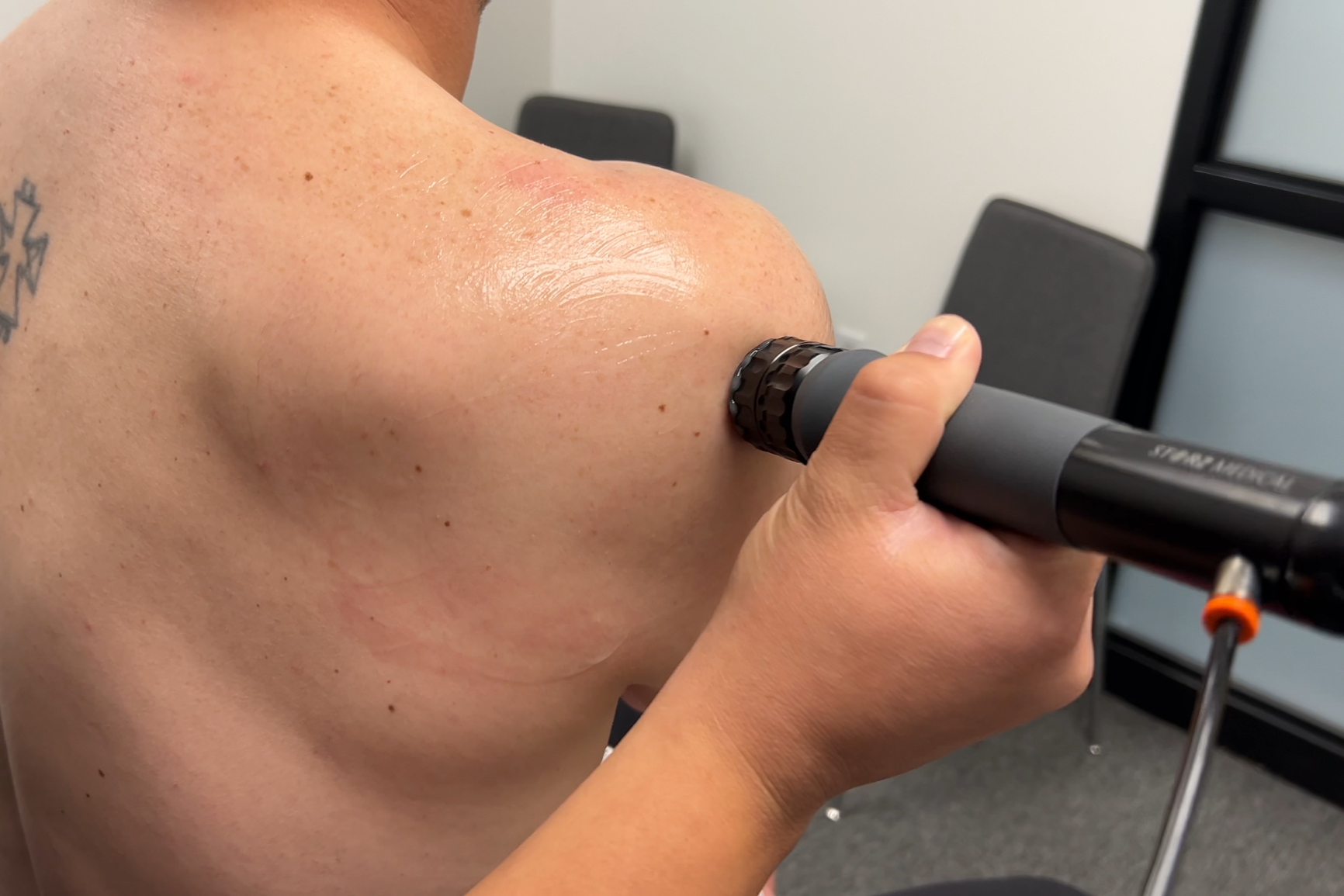
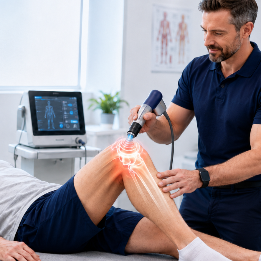
Shockwave therapy works by delivering controlled acoustic pulses through a handheld transducer into the injured area. A conductive gel is applied to the skin, and the device sends targeted sound waves that travel through the skin and soft tissue to reach the damaged tendon, fascia, or muscle. The mechanism of action is where the real science comes in. Here is what happens inside your body after a shockwave treatment:

1. Increased Blood Flow (Neovascularization)
The acoustic waves stimulate the formation of new blood vessels in the treated area. This process, called neovascularization, brings oxygen and nutrients to damaged tissues with poor blood supply, a key reason some tendon injuries fail to heal on their own. Research has shown that shockwave therapy enhances angiogenesis by activating VEGFR2, essentially signaling your body to build a new blood supply to the injury site.


2. Collagen Production and Tissue Remodeling
Shockwaves activate fibroblasts, the cells responsible for producing collagen. Over the weeks following treatment, your body works to reorganize disorganized scar tissue and lay down new, stronger collagen fibers in proper alignment. This is why shockwave therapy doesn't just reduce pain in the short term; it actually changes the tissue's structural integrity.


3. Pain Signal Disruption
Interestingly, many patients feel immediate pain relief after a session, even before the tissue has healed. This is due to something called the "analgesic effect," where the high-energy pulses temporarily disrupt pain signal transmission at the cellular level, giving patients relief while the deeper healing process gets underway.


4. Breakup of Calcific Deposits
For conditions like calcific tendinopathy of the shoulder, the acoustic energy can physically break up calcium deposits that have formed in the tendon, allowing the body to clear them naturally over time.


5. Anti-Inflammatory Modulation
Contrary to what you might expect, the initial response to shockwave therapy is actually pro-inflammatory. The treatment creates a controlled micro-trauma that restarts your body's healing process, recruiting immune cells, clearing out degenerated tissue, and signaling repair pathways. This is a key reason why you should NOT take anti-inflammatory medications in the 48 hours after treatment, as it would cut off the very process that the shockwave just activated.


Shockwave therapy works best when it is paired with the right rehab plan. The goal is not just to make the area feel better for a few days. The goal is to help the tissue become stronger, more resilient, and better prepared for running, lifting, walking, golf, hiking, or sports.

Conditions We Treat with Shockwave Therapy

Shockwave therapy is most effective for chronic tendon and soft tissue conditions, injuries that have been lingering for weeks or months and haven't responded to rest, stretching, or other conservative care. Here are some of the most common conditions we treat at our Murray, Utah, sports chiropractic clinic:


Plantar Fasciitis

This is the condition with the strongest evidence base for shockwave therapy. Plantar fasciitis affects the thick band of tissue on the bottom of the foot and can be notoriously stubborn to treat. Multiple studies and meta-analyses show shockwave therapy produces significant pain reduction in patients with chronic plantar fasciitis, often after just 2-3 sessions.


Achilles Tendinopathy

Whether you are a runner logging 40-mile weeks or a weekend warrior, Achilles pain can derail your entire season. Shockwave therapy targets degenerated tissue in the mid-portion of the tendon and stimulates new collagen formation, with studies showing that 60-80% of patients achieve good-to-excellent results.


Tennis Elbow / Golfer's Elbow (Lateral and Medial Epicondylitis)

These common overuse injuries of the elbow respond well to shockwave therapy, particularly when the condition has been present for more than three months. Repeated studies have shown that shockwave therapy is more effective than corticosteroid injections for long-term outcomes in lateral epicondylitis.


Patellar Tendinopathy (Jumper's Knee) and Runner’s Knee

A common issue for basketball, volleyball, and running athletes. Shockwave therapy has shown strong results in published systematic reviews, particularly when combined with eccentric loading exercises.


Rotator Cuff Tendinopathy / Shoulder Impingement

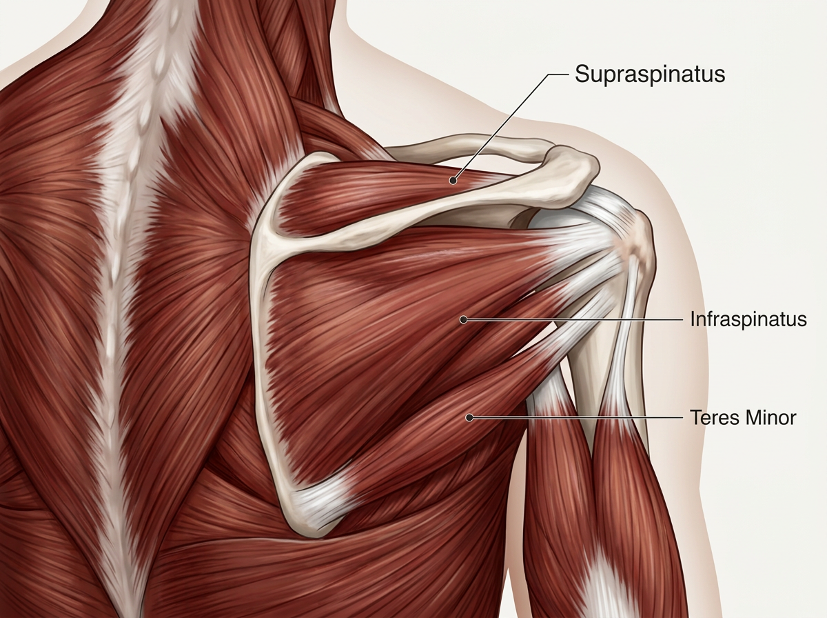
For shoulder pain not caused by a full-thickness tear, shockwave therapy can reduce pain and improve function by stimulating repair of the rotator cuff muscles and tendons.


Shin Splints (Medial Tibial Stress Syndrome), IT Band Syndrome, and Hamstring Tendinopathy

All of these conditions benefit from the increased blood flow and tissue remodeling that shockwave therapy promotes.


Shockwave therapy is not always the right treatment option. Acute tears, fractures, infections, certain nerve conditions, blood clotting issues, pregnancy, and other medical conditions may require a different approach. That is why we always start with a proper evaluation before recommending shockwave therapy.

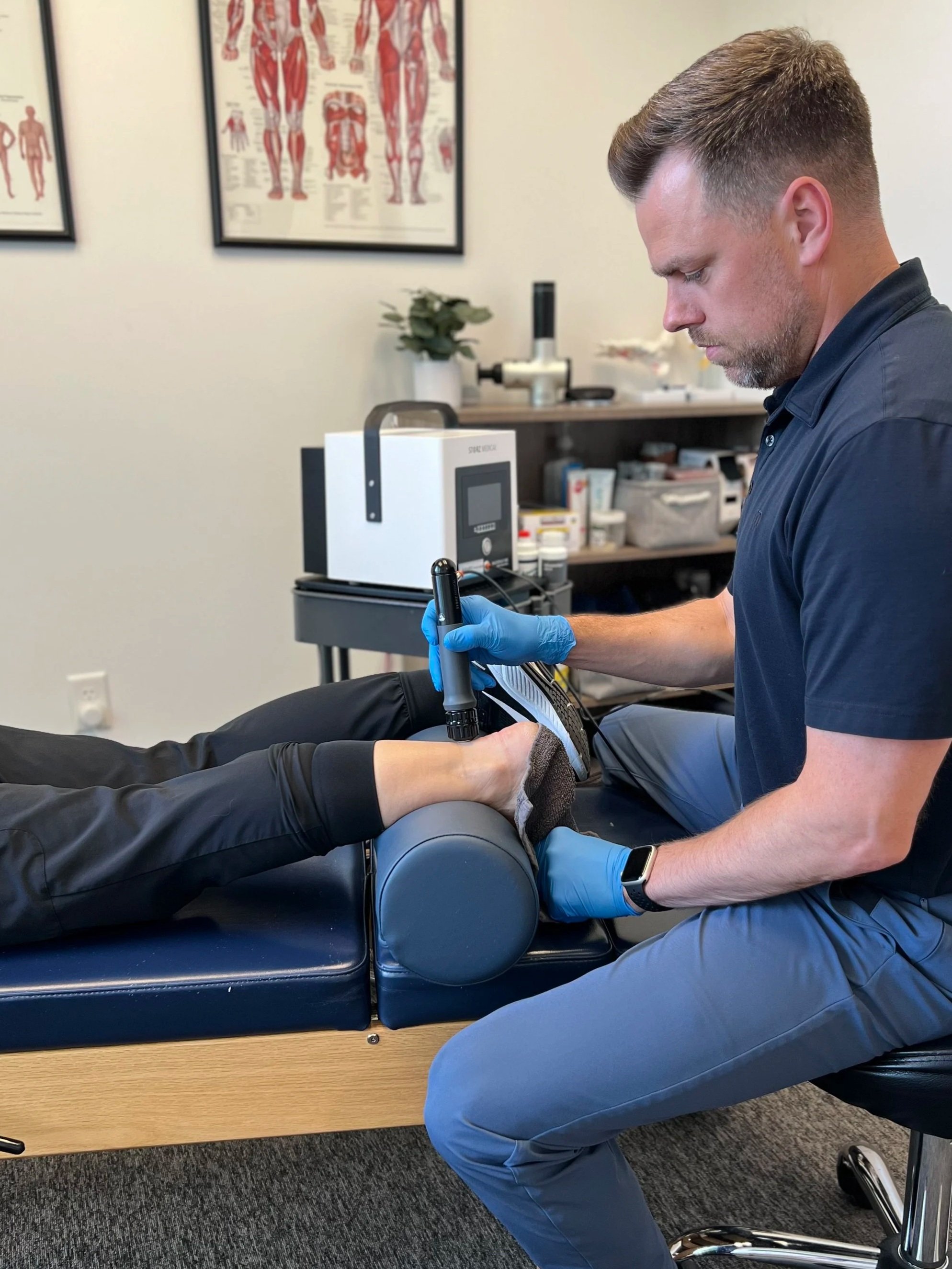
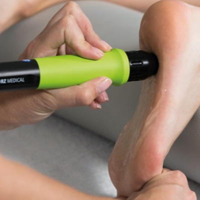
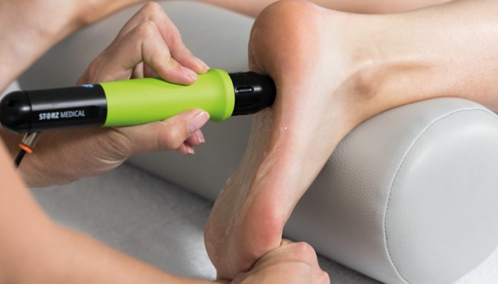
A chiropractor applying shockwave therapy to a patient's heel for plantar fasciitis treatment at Revive Sport & Spine in Murray, Utah.

What to Expect During Shockwave Treatment: Timeline and Soreness

One of the most common questions patients ask is, "Will it hurt?" Here is an honest answer and a realistic timeline so you know exactly what to expect.

During Treatment

  • A typical session lasts 5 to 15 minutes, depending on the area being treated. The sensation varies; some patients describe it as a deep, tapping pressure, while others feel brief, sharp pulses. The intensity is adjustable, and we work with you to find a level that is both effective and tolerable. No anesthesia or pain medication is needed.

Immediate Aftermath (0 to 48 Hours)

  • Many patients experience immediate pain relief due to the analgesic effect of the acoustic waves. However, as that wears off (usually within a few hours), you may feel a dull ache or soreness in the treated area, similar to the feeling after a heavy workout. Some mild swelling and tenderness are normal.

  • Critical: do NOT take NSAIDs for at least 48 hours after treatment unless directed to do so by your primary medical provider. These medications interfere with the inflammatory healing cascade that the shockwave just activated. Similarly, avoid icing the treated area; ice constricts blood flow and can blunt the healing response.

  • Light activity is fine, but avoid high-impact exercise or heavy lifting for 48 hours to let the tissue settle.

Weeks 1 to 4: The Cumulative Effect

  • Most patients need a series of 4-12 sessions, spaced 7-10 days apart. Healing is rarely a straight line; you may feel better after one session, sore after the next, then better again. This fluctuation is normal. Research shows that approximately 80% of patients report significant pain reduction after their third session.

  • Each session builds on the last, progressively breaking down damaged tissue and stimulating the production of healthy new tissue.

3 Months: Maximal Recovery

  • Here is what many patients don't realize: while you may feel significantly better within the first month, the biological remodeling of the tendon or fascia continues for several months. The tissue is literally rebuilding itself at the cellular level. Maximal recovery, where the tissue has fully matured and strengthened, typically occurs around the 12-week mark.

  • This is why we always pair shockwave therapy with corrective exercises and a rehab plan. The treatment creates the healing environment; the exercises retrain the tissue to handle the loads you need it to handle.


Why Choose Revive Sport & Spine for Shockwave Therapy in Murray?

At Revive Sport & Spine in Murray, UT, we use shockwave therapy for runners, golfers, lifters, youth athletes, and active adults who want to stay moving without relying only on rest, medication, or repeated short-term fixes.

Our approach is simple: find the source of the problem, treat the irritated tissue, build a plan around your goals, and help you return to the activities you enjoy.

If you are in Murray, Salt Lake City, Holladay, Midvale, Sandy, Cottonwood Heights, or Millcreek and have been dealing with chronic heel pain, Achilles pain, tendon pain, or a sports injury that will not go away, shockwave therapy may be worth considering.

Call Revive Sport & Spine at 801-944-1855 or schedule an appointment online to see if shockwave therapy is a good fit for your injury and your goals.

SOURCES:

  • Hatanaka, K., et al. "Molecular Mechanisms of the Angiogenic Effects of Low-Energy Shock Wave Therapy: Roles of Mechanotransduction." American Journal of Physiology-Cell Physiology, 2016. https://doi.org/10.1152/ajpcell.00152.2016

  • Molecular Medicine. "Extracorporeal Shock Wave Therapy Mechanisms in Musculoskeletal Regenerative Medicine." Journal of Clinical Orthopaedics and Trauma, vol. 11, Suppl. 3, 2020, pp. S309–S318. https://pmc.ncbi.nlm.nih.gov/articles/PMC7275282/

Injured in an Auto Accident? Chiropractic Care is an Important Part of your Recovery.

Auto accidents can cause a wide variety of injuries, and one of the most common types of injuries seen in these cases is whiplash. Whiplash is a neck injury that occurs when the head is suddenly and forcefully jerked forward and then backward, similar to the cracking of a whip. This type of injury can be caused by the sudden impact of a car accident, and it can cause a range of symptoms including neck pain, headaches, dizziness, and even tingling or numbness in the arms.

Another common injury seen in auto accidents is low back pain. The sudden impact of a car accident can cause the spine to be compressed, leading to strain and sprain of the muscles, ligaments, and even the bones of the lower back. This can cause a range of symptoms including pain, stiffness, and difficulty moving.

Chiropractic care can be an effective treatment option for both of these types of injuries. Chiropractors are trained to diagnose and treat a wide variety of musculoskeletal conditions, and they use a range of techniques to help alleviate pain and improve function.

When it comes to treating whiplash, chiropractors will often use manual techniques such as spinal manipulation or mobilization to help restore proper alignment to the spine and reduce pain and stiffness in the neck. They may also use other therapies such as deep tissue massage, ultrasound, or electrical stimulation to help relieve pain and inflammation. Chiropractors will also usually make recommendations for exercises and stretches that can help improve range of motion, reduce pain, and promote healing.

In the case of low back pain, chiropractors will also use manual techniques such as spinal manipulation or mobilization to help restore proper alignment to the spine and reduce pain and stiffness. They may also use other therapies such as deep tissue massage, ultrasound, or electrical stimulation to help relieve pain and inflammation. Chiropractors may also make recommendations for exercises and stretches that can help improve range of motion, reduce pain, and promote healing.

It's important to note that Chiropractors can also be helpful for the patient's rehabilitation process after a car accident by providing a proper diagnosis and treatment of their symptoms, as well as soft tissue therapies to help with any muscle spasms, strains or sprains.

Additionally, Chiropractors may also collaborate with other healthcare providers such as primary care physicians, physical therapists, or specialists, if necessary. They may refer patients to these providers for additional testing or treatment, or they may work with these providers as part of a multidisciplinary team to provide the best possible care for the patient.

In conclusion, chiropractic care can be an effective treatment option for both whiplash and low back pain caused by auto accidents. Chiropractors use a variety of techniques to alleviate pain and improve function, and they can also play an important role in the rehabilitation process after a car accident. Additionally, working with other healthcare providers as needed, making chiropractic care a very valuable asset in the healing process after an auto accident

What Causes Sciatica and How Sports Chiropractic Care Can Help?

What Causes Sciatica and How Sports Chiropractic Care Can Help?

The sciatic nerve runs from the lower back through each buttock and down each leg. It’s the largest nerve in the body and helps us with stability and movement, making it significant to many of our everyday activities.

How Chiropractic Care Can Alleviate Sciatica

How Chiropractic Care Can Alleviate Sciatica

Millions of Americans suffer from sciatica every year; sometimes, the pain is minimal, and other times it’s intense. In either case, finding the right chiropractic treatment will ensure you get optimized care that delivers results.


Chiropractic techniques are proven to end pain by finding what’s causing it. This comprehensive approach will dictate a wellness plan that reestablishes and restores the body’s full function, taking pressure off affected areas, including those found within the sciatic nerve.


5 Areas To Develop Great Habits That Will Improve Your Health

5 Areas To Develop Great Habits That Will Improve Your Health

With the continuation and exacerbated condition of the COVID-19 pandemic, this might be a challenging question to answer. You may feel physically well. Heck, you may even be one of the strong ones, unphased by the "stay home" orders and are socially and mentally doing well also.

Regardless of how you respond to the initial question, I have one response for you.

New Year - New Goals: Setting & Achieving Your Goals This Year.

New Year - New Goals:  Setting & Achieving Your Goals This Year.

If you are like me, you love to set goals, spend hours on action plans to achieve those goals, and ponder on the impact and improvements you can make to yourself, your company, your family, your health, and everything else around you.